

Laboratories & Physicians
Epidemiology
Dermatophytosis: a widespread and evolving challenge
Dermatophyte infections occur worldwide, with patterns shaped by age, sex, climate
and geography.
The epidemiological picture, is far from uniform:
20–25%
Estimated global prevalence of superficial fungal infections¹
Age matters
Tinea capitis mainly affects children, while nail and foot infections are more frequent in adults.²˒³
Sex patterns vary
Male predominance is particularly reported for tinea pedis, although patterns differ between clinical forms and populations.²
Dermatophyte epidemiology continues to change, with shifts in circulating species,
population groups affected and antifungal resistance creating new diagnostic challenges.
Explore three key epidemiological stories shaping dermatophyte diagnostics.
-
Tinea Capitis
A Paediatric Challenge
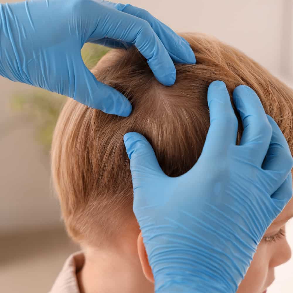
Tinea capitis mainly affects children and can cause:
- Scaling
- Itching
- Hair breakage or hair loss
- Inflammatory lesions in some cases
- Transmission between children may contribute to school or household outbreaks4.
Why diagnosis matters:
Clinical presentation can vary, while scalp involvement generally requires a different management approach from localized skin disease.
-
Onychomycosis
Persistent by Nature
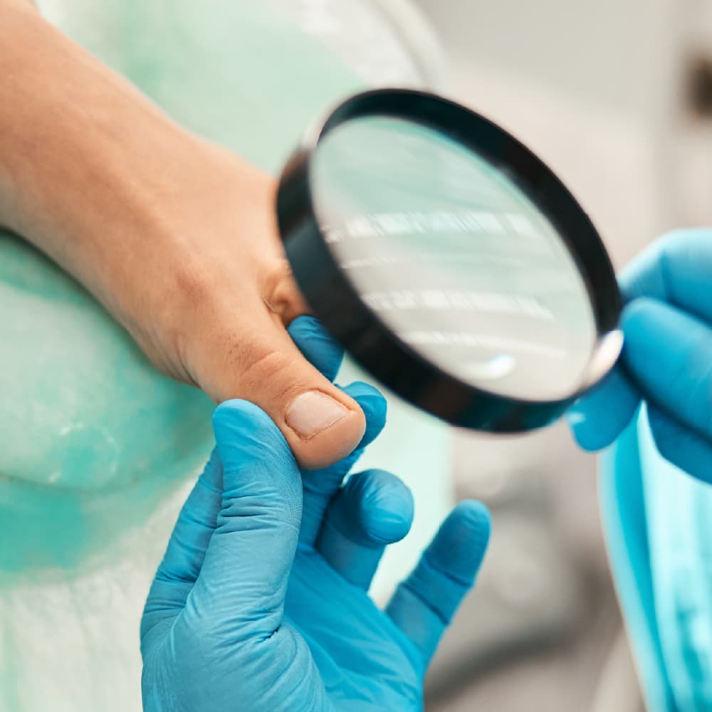
Nail dermatophytosis can present with:
- Thickening
- Discoloration
- Brittleness
- Subungual debris
However, not every abnormal nail is fungal.
Current guidance therefore supports mycological confirmation using microscopy, culture and/or molecular methods as part of the diagnostic work-up 5.
A diagnostic challenge
Nail specimens can be difficult to analyse, and fungal culture may require up to approximately four weeks of incubation.
-
Trichophyton indotineae
An Emerging European Challenge
Why is it important?
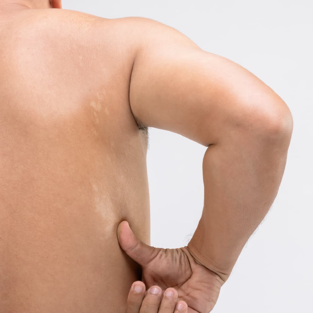
- Can cause extensive dermatophytosis
- May be associated with recurrent or difficult-to-treat disease
- Terbinafine resistance has been increasingly reported associated with mutations in squalene epoxidase (SQLE) gene6 *
*A systematic review of European reports identified 63 antifungal-resistant cases
from 16 studies published between 2019 and 20236.
A growing diagnostic need
Emerging species and resistance patterns increase the value of accurate organism identification.
From epidemiology to diagnosis
Understanding who is affected is only part of the picture. Explore how dermatophyte infections are identified in the laboratory and which diagnostic methods can be used.
References
- Kovitwanichkanont T, Chong AH. Cutaneous fungal infections caused by dermatophytes and non-dermatophytes: an updated comprehensive review of epidemiology, clinical presentations, and diagnostic testing. J Fungi (Basel). 2023;9(6):669. doi:10.3390/jof9060669.
- Leung AKC, Barankin B, Lam JM, Leong KF, Hon KL. Tinea pedis: an updated review. Drugs Context. 2023;12:2023-5-1. doi:10.7573/dic.2023-5-1.
- Sigurgeirsson B, Baran R. The prevalence of onychomycosis in the global population: a literature study. J Eur Acad Dermatol Venereol. 2014;28(11):1480–1491. doi:10.1111/jdv.12323.
- World Health Organization. Ringworm (tinea). WHO; 19 June 2025.
- Nenoff P, Reinel D, Mayser P, et al. S1 Guideline onychomycosis. J Dtsch Dermatol Ges. 2023;21(6):678– 692. doi:10.1111/ddg.14988.
- Ferreira CB, Lisboa C. A systematic review on the emergence of terbinafine-resistant Trichophyton indotineae in Europe: Time to act? J Eur Acad Dermatol Venereol. 2025;39(2):364–376. doi:10.1111/jdv.20270.