

Eurobio Scientific
Diagnostics
Diagnostic Objectives
The primary objectives in the diagnostic work-up of suspected dermatophyte infections are to:
- Confirm fungal infection and distinguish it from other dermatological conditions.
- Identify the causative organism where clinically relevant.
- Support appropriate clinical management, particularly in persistent, recurrent or atypical disease.
- Support differential diagnosis when laboratory findings do not confirm dermatophytosis.1–3
Accurate diagnosis combines clinical evaluation, appropriate sampling and laboratory investigation.1,2

Eurobio Scientific
Diagnostic Approach
Clinical suspicion
Appropriate sampling
Laboratory investigation
Direct Microscopy
Rapid visualization of fungal elements
Clinical material is commonly treated with a clearing agent such as potassium hydroxide (KOH) to facilitate visualization of fungal structures.
Strengths
- Rapid
- Relatively simple
- Provides direct evidence of fungal elements
Limitations
- Operator dependent
- Strongly influenced by sample quality
- Limited ability to identify the causative species 1,2
Fungal Culture
Isolation of viable fungi
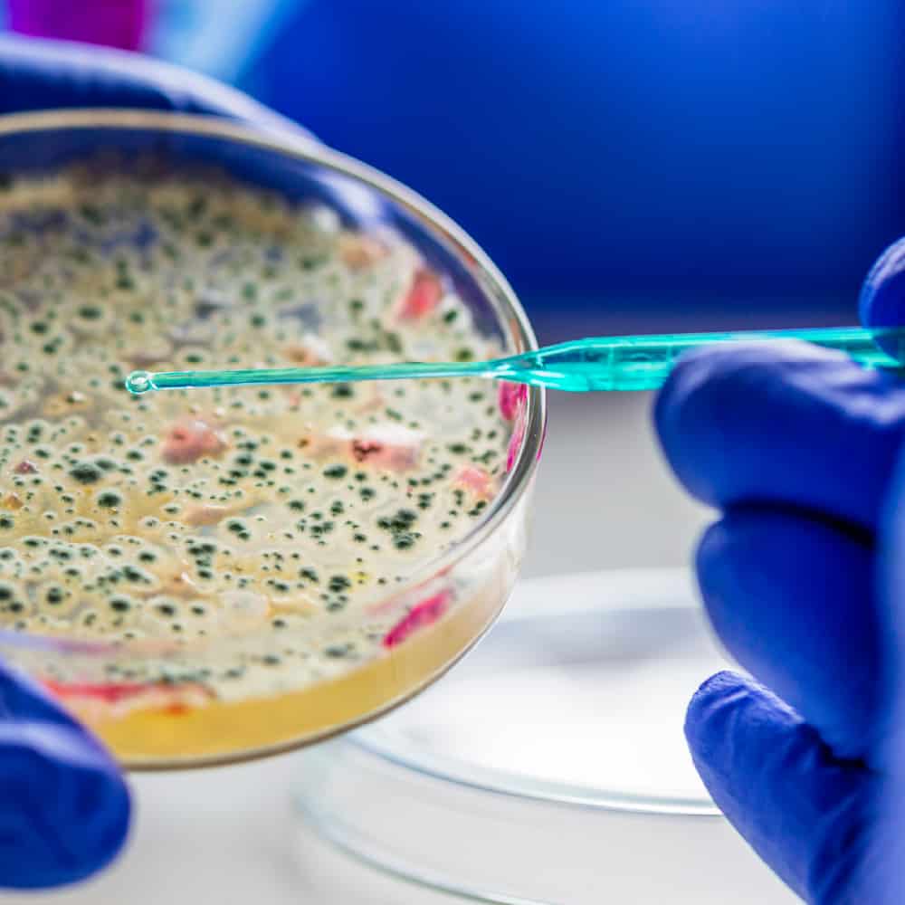
Specimens are inoculated onto suitable mycological media to allow fungal growth and subsequent identification.
Strengths
- Isolation of viable fungi
- Can support species identification
- Provides isolates for further investigation
Limitations
- Long turnaround time
- Variable diagnostic sensitivity
- Potential contamination
- Requires morphological expertise1,2
For nail specimens, the S1 guideline recommends approximately 4 weeks of incubation for dermatophyte culture.3
Molecular Diagnosis
PCR detection of fungal DNA
PCR-based methods detect predefined fungal DNA targets directly from clinical specimens, without requiring fungal growth.
Potential advantages
- Rapid detection*
- Direct testing from the clinical specimen
- Multiplex identification of several targets may be possible
- Reduced dependence on fungal morphology
- Does not require viable fungi for detection
Unlike culture, PCR does not rely on fungal viability. Detection may therefore remain possible when antifungal treatment has already been started, although results should always be interpreted in the clinical context.

References
- Kidd SE, Weldhagen GF. Diagnosis of dermatophytes: from microscopy to direct PCR. Microbiology Australia. 2022;43(1):9–13. doi:10.1071/MA22005.
- Aho-Laukkanen E, Mäki-Koivisto V, Torvikoski J, Sinikumpu SP, Huilaja L, Junttila IS. PCR enables rapid detection of dermatophytes in practice. Microbiology Spectrum. 2024;12(11):e01049-24. doi:10.1128/spectrum.01049-24.
- Nenoff P, Reinel D, Mayser P, et al. S1 Guideline onychomycosis. J Dtsch Dermatol Ges. 2023;21(6):678–692. doi:10.1111/ddg.14988.